

Ripla Arora
 Contact
ripla@msu.edu
517-353-6958
View publications on MSU Scholars
IQ DIVISION – Developmental and Stem Cell Biology
Departmental AFFILIATIONS
About
Ripla Arora is an associate professor in the Department of Obstetrics, Gynecology, and Reproductive Biology within the College of Human Medicine. Her research focuses on embryo uterine interactions at the time of implantation and uterine development.
Dr. Arora received her undergraduate and graduate degrees in biochemistry from the University of Delhi, India. She went on to receive her Ph.D. degree in genetics and development from Columbia University, completing her postdoctoral work in the Department of Obstetrics Gynecology and Reproductive Sciences at the University of California-San Francisco.
The Arora Lab
Early events of mammalian pregnancy, although crucial for normal development, are poorly understood. Owing to the small size and transparent nature of the early embryo, development from fertilization to blastocyst stages has been extensively documented by imaging. On the other hand, a deeper understanding of the 3D architecture of the embryo’s uterine environment and the uterine glands has lagged behind.
The Arora Lab uses developmental genetics, 3D imaging, computational image analysis, and gene expression analysis, to understand how hormones influence the uterine architecture to modulate receptivity and implantation.
Main projects
Hormonal Regulation of Receptivity and Implantation
The uterine tube folds randomly in the absence of pregnancy but folds in a stereotypic manner to create uterine crypts that home the incoming embryos. We are using genetic mouse mutants, gene expression analysis and 3D imaging to understand how hormones guide the formation of the crypts.
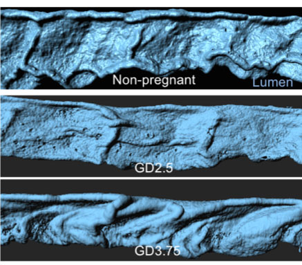
Dynamic folding of the mouse uterine lumen (blue) at different days of mouse pregnancy.
Uterine Gland Branching Morphogenesis
In a mammalian pregnancy, uterine glands nourish the embryo until the placenta forms. The uterine glands undergo patterning and reorganization at the time of embryo implantation. Using different image analysis platforms we are dissecting the structure of the uterine glands and the changes that accompany implantation. Using spatial localization of the glands with respect to the other cell types, we will determine the functional consequences of these architectural glandular changes.
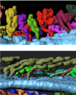
Uterine glands depicted by pseudocolors connected to the lumen (blue) in the absence (top) and the presence of an embryo (bottom).
Quantitative Imaging and Modeling
We are using quantitative approaches to measure changes that occur in the uterine luminal and glandular architecture to prepare for pregnancy. We are also applying these methods to conditions where implantation fails to occur
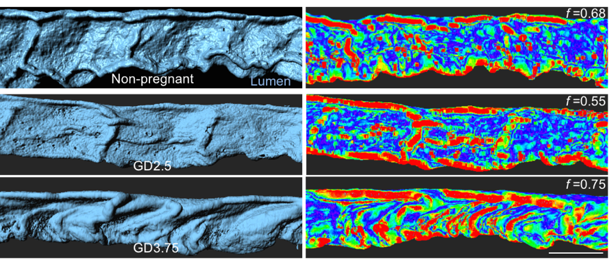
Quantification (right) of folding patterns (left) using matlab, depicted as a heat map.