

Jonathan Hardy
 Contact
hardyjon@msu.edu
Office: 517-884-6971
Lab: 517-355-4067
View publications on MSU Scholars
IQ DIVISION – Developmental and Stem Cell Biology
Departmental AFFILIATIONS
About
Jonathan Hardy is an associate professor in MSU’s Department of Microbiology and Molecular Genetics. He was recruited from Stanford to join MSU’s Institute for Quantitative Health Science & Engineering. Dr. Hardy has a B.S. in cell and molecular biology from the University of Washington and a Ph.D. in microbiology and immunology from Stanford University.
The Hardy Lab
The Hardy Laboratory uses the techniques of molecular imaging to investigate the mechanisms and consequences of bacterial infections that affect children. We are interested in prenatal and perinatal infections, as well as lung, wound, and implant infections that involve bacterial biofilms. We are also interested in oral bacteria that can cause preterm birth. We are researching novel treatments including plant products, bacteriophage, and antibiotic synergy. The modalities of molecular imaging allow us to localize and quantify bacterial infection in live animals over time, non-invasively.

Research
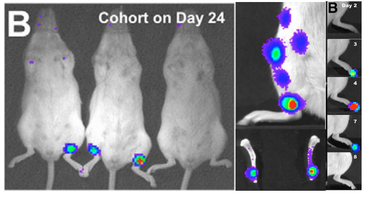
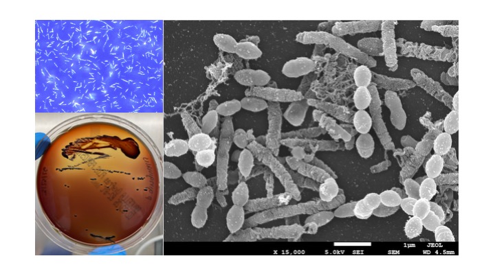
Using in vivo bioluminescence imaging (BLI), we discovered that Listeria monocytogenes, a deadly foodborne bacterial pathogen, replicates extracellularly in the lumen of the gallbladder. This normally intracellular pathogen grows to very high numbers in this organ, often to higher overall numbers than anywhere else in the body. This growth pattern was later confirmed to occur in humans and could represent an asymptomatic carrier state similar to that of typhoid fever.
Extracellular replication of Listeria monocytogenes in the murine gall bladder. Science. 2004 Feb 6;303(5659):851-3. doi: 10.1126/science.1092712. PMID: 14764883
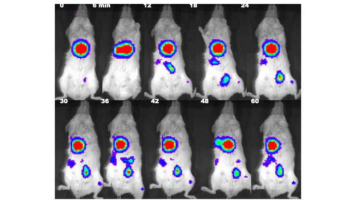
Excretion of Listeria from the gallbladder can be imaged using BLI. When a mouse with Listeria in the gallbladder is fed milk to cause the organ to contract, BLI signals from the ejected bacteria move though the intestine of the animal and can be followed with sequential imaging.
Induced biliary excretion of Listeria monocytogenes. Infect Immun. 2006 Mar;74(3):1819-27. doi: 10.1128/IAI.74.3.1819-1827.2006. PMID: 16495556
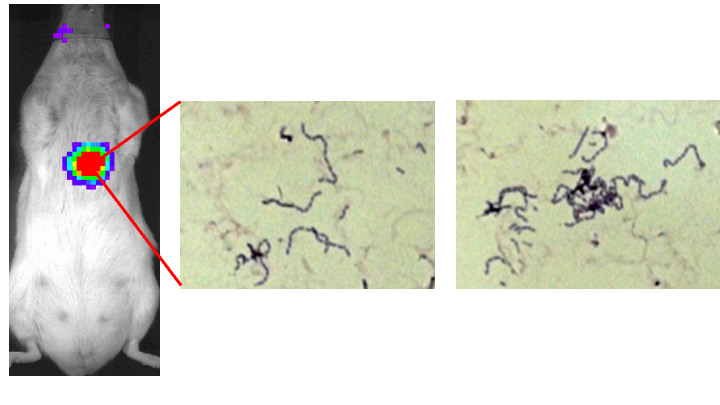
Listeria can persist for weeks in the bone marrow. It hides there without causing any outward symptoms or histological evidence. BLI revealed the bone marrow as a previously unsuspected location of growth of this pathogen.
Foci of Listeria monocytogenes persist in the bone marrow. Dis Model Mech. 2009 Jan-Feb;2(1-2):39-46. doi: 10.1242/dmm.000836. Epub 2008 Dec 22. PMID: 19132117
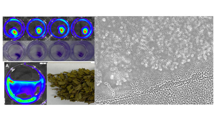
Bacterial biofilms are aggregates of bacterial cells adhering to a surface. The bacteria live within an extracellular matrix (ECM) composed of polysaccharide, protein, and DNA. Biofilms can form in lung infections such as those that occur in children with cystic fibrosis (CF). The formation of biofilms in the lungs of CF patients makes the bacteria more resistant to antibiotic treatment. The ECM may shield the bacteria from antibiotics, and some of the bacteria within the biofilm alter their metabolism, making them more resistant. The Hardy Lab is investigating novel antibiofilm treatments, including plant products such as cinnamaldehyde and extracts of the plant Larrea tridentata. The effects can be monitored using BLI. We are also testing therapies based on bacterophages, which are viruses that kill bacteria, in collaboration with the Eliava Institute of Bacteriophages, Microbiology and Virology in Tbilisi, Republic of Georgia.
Biofilm and planktonic Staphylococcus aureus exhibit distinct gene expression patterns in response to cinnamaldehyde. Infect Genet Evol. 2026 Apr;139:105919. doi: 10.1016/j.meegid.2026.105919. Epub 2026 Mar 4. PMID: 41791499
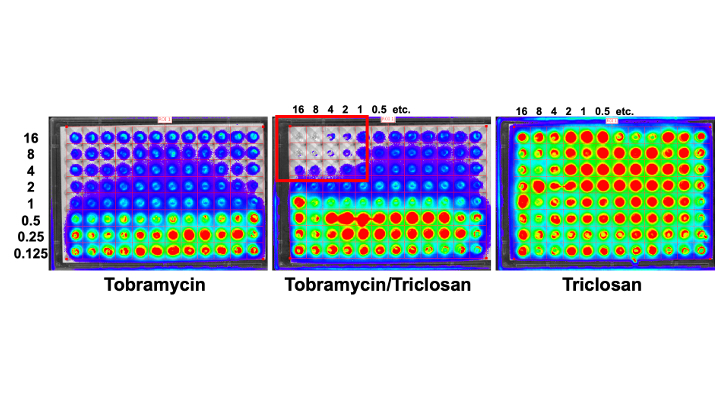
One improvement for the use of antibiotics against biofilm infections is antibiotic synergy. Two antibiotics or an antibiotic and another substance can increase the efficiacy of the treatment. We use bioluminescence as a tool for imaging biofilm responses of Pseudomonas aeruginosa, which infects CF patients, to individual and combined antibiotic treatments.
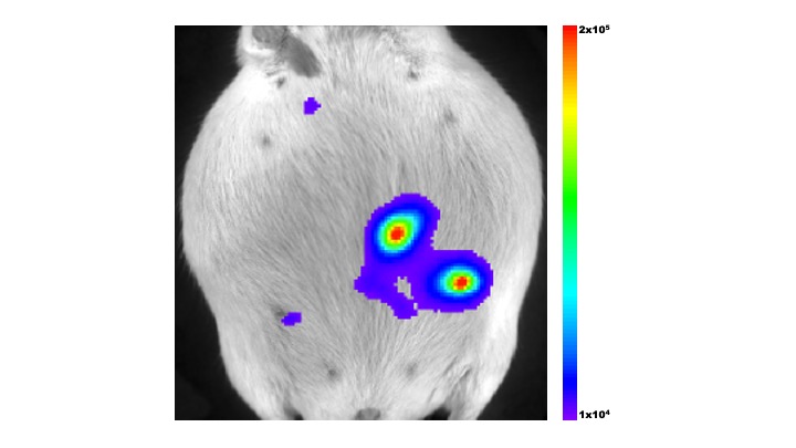
Prenatal infection by several bacterial pathogens results in a wide range of consequences to the fetus. Maternal and fetal responses to bacterial infection are poorly understood, partly because of a complex balance of immunity and tolerance. We use the pregnant mouse model of listeriosis to study the mechanisms and consequences of prenatal infection. Listeria infects the placenta, the incredible interface between mother and fetus. BLI can localize and quantify placental infection by Listeria.
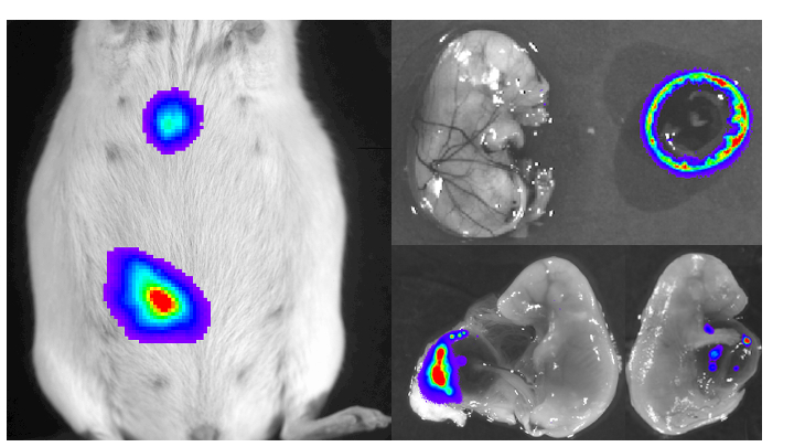
BLI permits the localization of bacterial infection in excised tissues, providing a powerful method of image-guided analysis. By controlling the dose of Listeria in pregnant mice and imaging the excised tisssues, we can analyze the effects of placental infections that have not crossed into the fetus itself. One of the targets of Listeria infection in the placenta are fetal trophoblast cells.
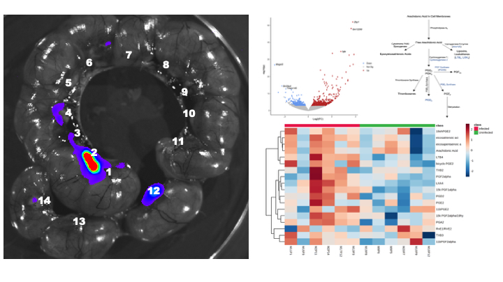
Image-guided analysis is used to compare infected and uninfected placentas from the same pregnant mouse, thereby controlling for systemic factors such as immune responses in the maternal blood. Analysis using this method revealed changes in the eicosanoid pathway that produces prostaglandins and leukotrienes that regulate development, pregnancy, and immune responses. Gene expression studies identified key enzymes such as Ido1 that are induced by placental infection and have multiple effects on fetal development.
Infection with Listeria monocytogenes alters the placental transcriptome and eicosanome. Placenta. 2022 Oct;128:29-35. doi: 10.1016/j.placenta.2022.08.001. Epub 2022 Aug 23. PMID: 36057170
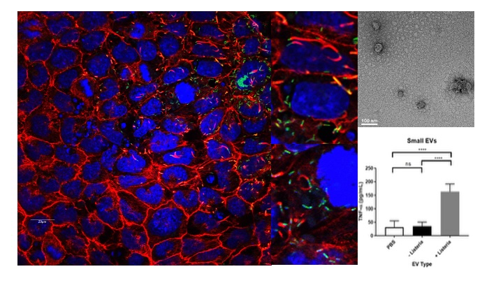
Trophoblasts, one of the most amazing cell types in all of nature, are fetal cells in the placenta that are in direct contact with maternal blood. They protect the fetus from maternal immune responses while permitting the exchange of nutrients, crucial hormones, and many other factors necessary for fetal development. Extracellular vesicles (EVs) are tiny membrane-bound particles produced by all cells of the body. Fetal trophoblast EVs flood the maternal blood during pregnancy and regulate the maternal immune system. However, the mechanisms of this process are very poorly understood. Infection by Listeria alters trophoblast EVs making them capable of immune stimulation. Infection loads the EVs with damage associated molecular patterns (DAMPs), but there are surprisingly no bacterial products in the EVs at all. DAMPs may be a major mechanism of immune regulation by trophoblasts during pregnancy, possibly regulating physiological disorders such as preeclampsia.
Listeria monocytogenes Infection Alters the Content and Function of Extracellular Vesicles Produced by Trophoblast Stem Cells. Infect Immun. 2022 Oct 20;90(10):e0034722. doi: 10.1128/iai.00347-22. Epub 2022 Sep 26. PMID: 36154271
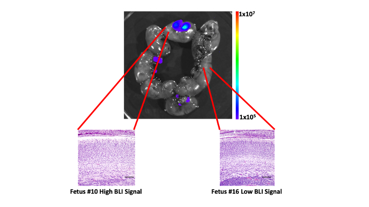
Placental infection alters fetal brain development. BLI is used to identify infected placentas and compare the associated fetal brains to uninfected fetuses in the same pregnant animal. In cases of significant infection, fetal brain layering is altered. These layers are formed by migration of neurons in the brain. Even subtle alterations of neuronal distribution patterns can result in neurodevelopmental conditions such as schizophrenia, bipolar disorder, and autism. Prenatal infection models in mice may reveal specific mechanisms of these disorders in humans.
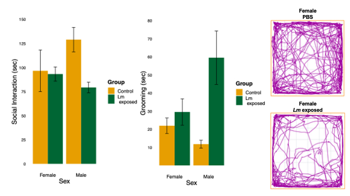
Offspring of mice infected during pregnancy can appear outwardly normal in every way but exhibit altered behavior. Changes in fetal brain development due to placental infection can result in sex-specific behavioral effects. Female and male offspring show distinct behavioral alterations due to prenatal exposure to Listeria. Sex-specific differences in social interaction, grooming, and open field exploration are indicative of neurodevelopmental changes that resemble human autism.
Infection of the murine placenta by Listeria monocytogenes induces sex-specific responses in the fetal brain. Pediatr Res. 2023 May;93(6):1566-1573. doi: 10.1038/s41390-022-02307-1. Epub 2022 Sep 20. PMID: 36127406
Bacteria that live in the oral cavity can escape to other tissues of the body and cause disease. The dental bacteria Fusobacterium nucleatum and Porphyromonas gingivalis that inhabit the gingival space between the tooth and gum are known to be released from periodontal infection sites into the bloodstream. If they access the placenta, they can cause preterm birth. We are interested in how these bacteria interact in anaerobic biofilms and how they cause preterm labor. We hope to one day image the processes with BLI, and are trying to engineer these bacteria to produce light.